Cell Structure (Cambridge International IGCSE Biology)
Animal & plant cells
Spec point —
2.1.1· Describe and compare the structure of a plant cell with an animal cell, limited to: cell wall, cell membrane, nucleus, cytoplasm, chloroplasts, ribosomes, mitochondria, vacuoles
Animal cell structure
-
The main features of animal cells:
- they contain a nucleus with a distinct membrane
- cells do not have cellulose cell walls
- their cells do not contain chloroplasts (so they are unable to carry out photosynthesis)
- they contain carbohydrates stored as glycogen
Animal cell diagram
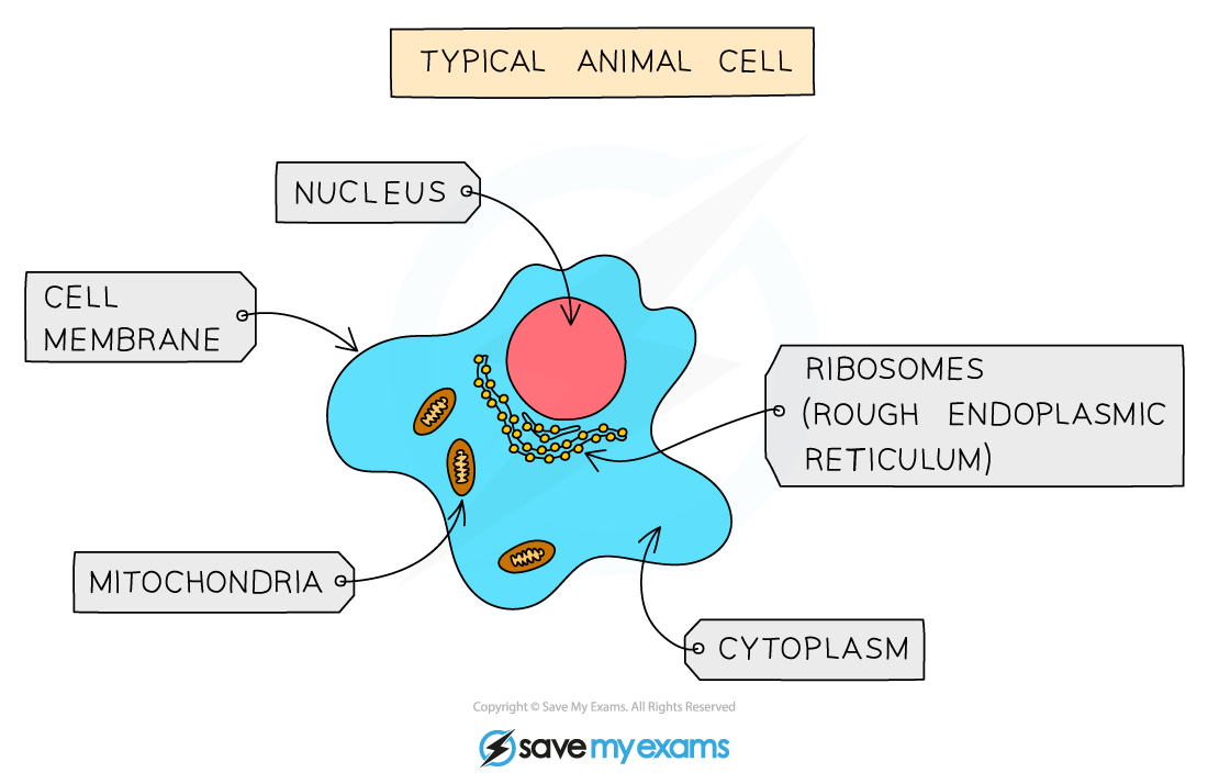
A typical animal cell
Plant cell structure
-
The main features of plant cells:
- they contain a nucleus with a distinct membrane
- cells have cell walls made out of cellulose
- they contain chloroplasts (so they can carry out photosynthesis)
- carbohydrates are stored as starch or sucrose
Plant cell diagram
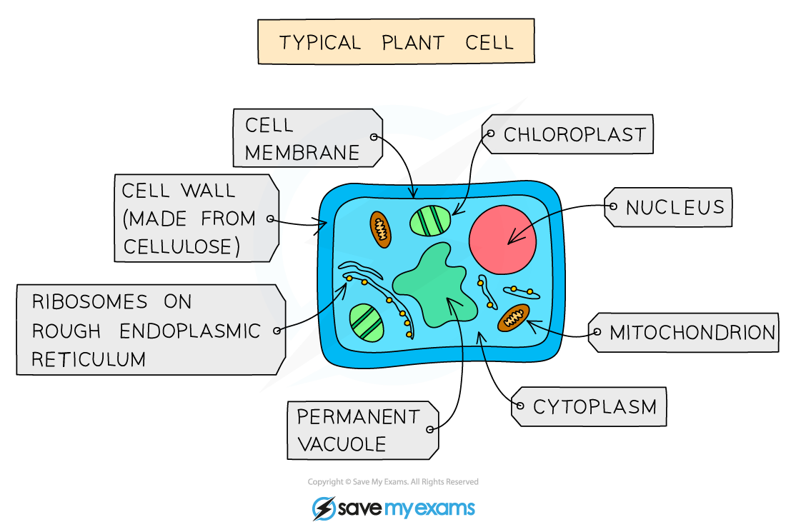
A typical plant cell
Animal and plant cell structure and function table
| Structure | Function |
|---|---|
| Nucleus | - Contains the DNA (genetic material) which controls the activities of the cell |
| Cytoplasm | - A gel like substance composed of water and dissolved solutes - Supports the internal structures of the cell - Site of many chemical reactions (including anaerobic respiration) |
| Cell membrane | - Holds the cell together separating the inside of the cell from the outside - Controls which substances enter or leave the cell |
| Ribosomes | - Found in the cytoplasm - The site of protein synthesis |
| Mitochondria | - The site of aerobic respiration |
Cell structure diagram
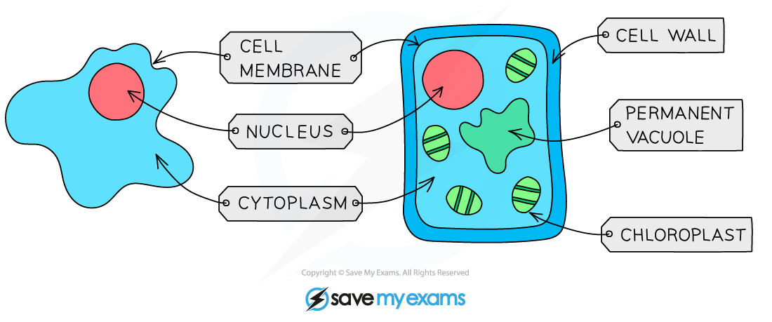
An animal and plant cell as seen under a light microscope
Additional plant cell structure and function table
| Structure | Function |
|---|---|
| Cell wall | - Made of cellulose(a polymer of glucose) - Gives the cell extra support, defining its shape |
| Chloroplast | - Contains the green chlorophyll pigment that absorbs light energy for photosynthesis |
| Permanent vacuole | - Contains cell sap: a solution of sugar and salt - Used for storage of certain materials - Helps to support the shape of the cell |
Bacteria cells
Spec point —
2.1.1· Describe the structure of a bacterial cell, limited to: cell wall, cell membrane, cytoplasm, ribosomes, circular DNA, plasmids
Bacteria cell structure
-
Bacteria, which have a wide variety of shapes and sizes, all share the following biological characteristics:
- they are microscopic single-celled organisms
- possess a cell wall (made of peptidoglycan, not cellulose), cell membrane, cytoplasm and ribosomes
- lack a nucleus but contain a circular chromosome of DNA that floats in the cytoplasm
- plasmids are sometimes present - these are small rings of DNA (also floating in the cytoplasm) that contain extra genes to those found in the chromosomal DNA
- they lack mitochondria, chloroplasts and other membrane-bound organelles found in animal and plant cells
-
Some bacteria also have a flagellum (singular) or several flagella (plural). These are long, thin, whip-like tails attached to bacteria that allow them to move
-
Examples of bacteria include:
- Lactobacillus (a rod-shaped bacterium used in the production of yoghurt from milk)
- Pneumococcus (a spherical bacterium that acts as the pathogen causing pneumonia)
Bacteria cell diagram
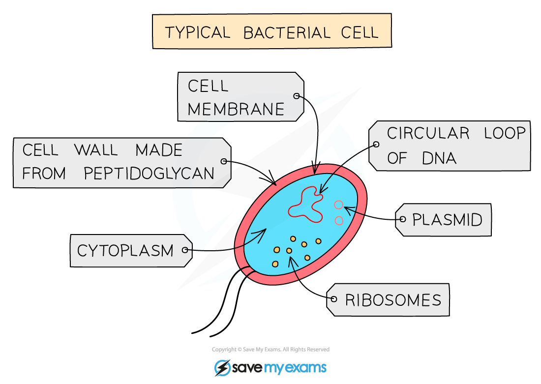
A typical bacterial cell
Identifying cell structures & function
Spec point —
2.1.1· Identify the cell structures listed in diagrams and images of plant, animal and bacterial cells Describe the functions of the structures in plant, animal and bacterial cells
Identifying cell structures & function
-
Within the cytoplasm, the following organelles are visible in almost all cells except prokaryotes when looking at higher magnification (ie using an electron microscope):
- mitochondria (singular: mitochondrion) are organelles found throughout the cytoplasm
- ribosomes are tiny structures that can be free within the cytoplasm or attached to a system of membranes within the cell known as endoplasmic reticulum
- Endoplasmic reticulum studded with ribosomes looks rough under the microscope; this gives rise to its name of rough endoplasmic reticulum (often shortened to R.E.R.)
- vesicles can also be seen using a higher magnification - these are small circular structures found moving throughout the cytoplasm
Identifying cell structures under a microscope
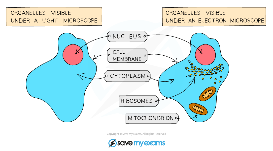
Structures in an animal cell visible under a light microscope and an electron microscope
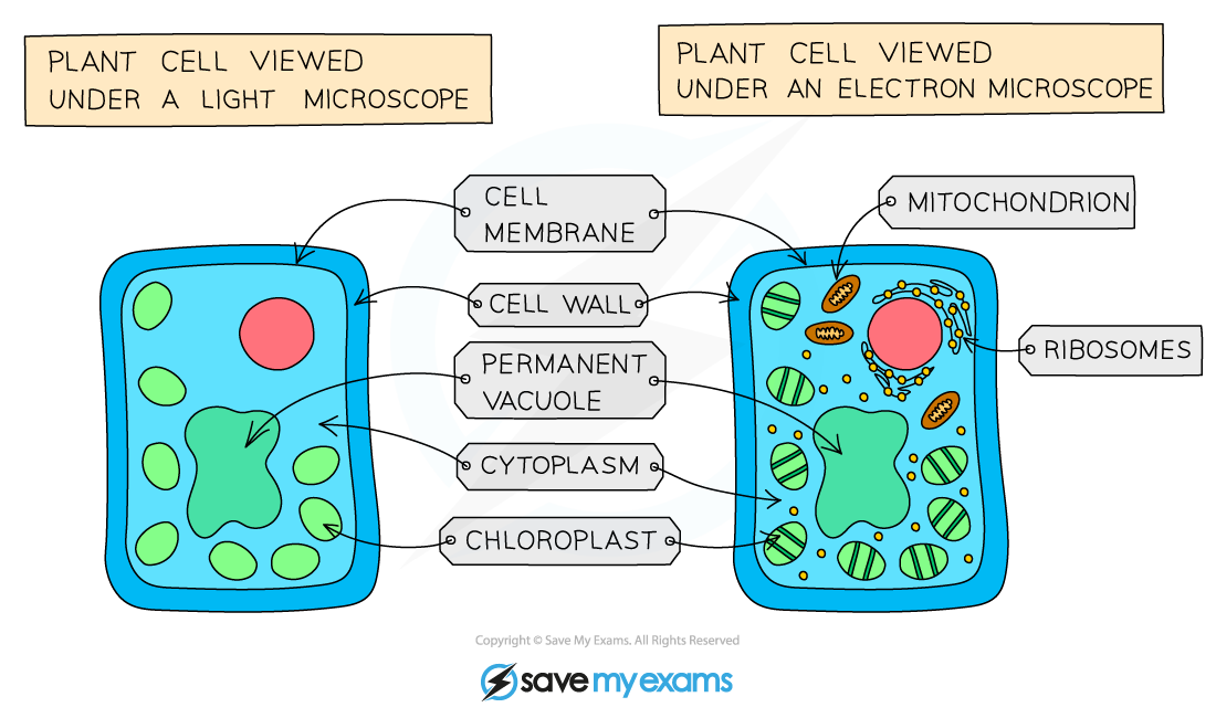
Structures in a plant cell visible under a light microscope and an electron microscope
Was this revision note helpful?
Build on this topic