Kidney Structure (Cambridge International A-Level Biology)
Structure of the Human Kidney
Spec point —
14.1.4· describe the structure of the human kidney, limited to: • fibrous capsule • cortex • medulla • renal pelvis • ureter • branches of the renal artery and renal vein
Structure of the human kidney
-
Humans have two kidneys
-
The kidneys are responsible for carrying out two very important functions:
- As an osmoregulatory organ—they regulate the water content of the blood (vital for maintaining blood pressure)
- As an excretory organ—they excrete the toxic waste products of metabolism (such as urea) and substances in excess of requirements (such as salts)
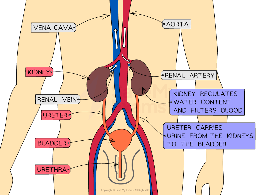
The position of the kidneys and their associated structures
| Structure | Function |
|---|---|
| Renal artery | Carries oxygenated blood (containing urea and salts) to the kidneys |
| Renal vein | Carries deoxygenated blood (that has had urea and excess salts removed) away from the kidneys |
| Kidney | Regulates water content of blood and filters blood |
| Ureter | Carries urine from the kidneys to the bladder |
| Bladder | Stores urine (temporarily) |
| Urethra | Releases urine outside of the body |
-
The kidney itself is surrounded by a fairly tough outer layer known as the fibrous capsule
-
Beneath the fibrous capsule, the kidney has three main areas:
- The cortex (contains the glomerulus, Bowman’s capsule, proximal convoluted tubule, and distal convoluted tubule of the nephrons)
- The medulla (contains the loop of Henle and collecting duct of the nephrons)
- The renal pelvis (where the ureter joins the kidney)
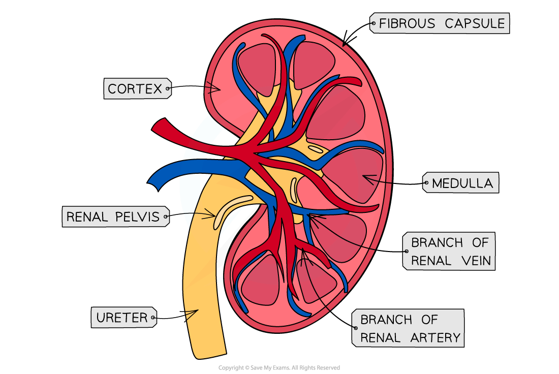
A cross-section of a kidney (as seen when cut in half vertically)
Exam Hint Make sure you can identify all the structures mentioned on this page if you are presented with a diagram of the kidney and its associated structures, or a diagram with a vertical cross-section of the kidney itself.
Was this revision note helpful?
Build on this topic