Ultrastructure of Striated Muscle (Cambridge International A-Level Biology)
Spec point —
15.1.11· describe the ultrastructure of striated muscle with reference to sarcomere structure using electron micrographs and diagrams
Ultrastructure of striated muscle
-
Striated muscle makes up the muscles in the body that are attached to the skeleton
- 'Striated' means it is striped/streaky in appearance
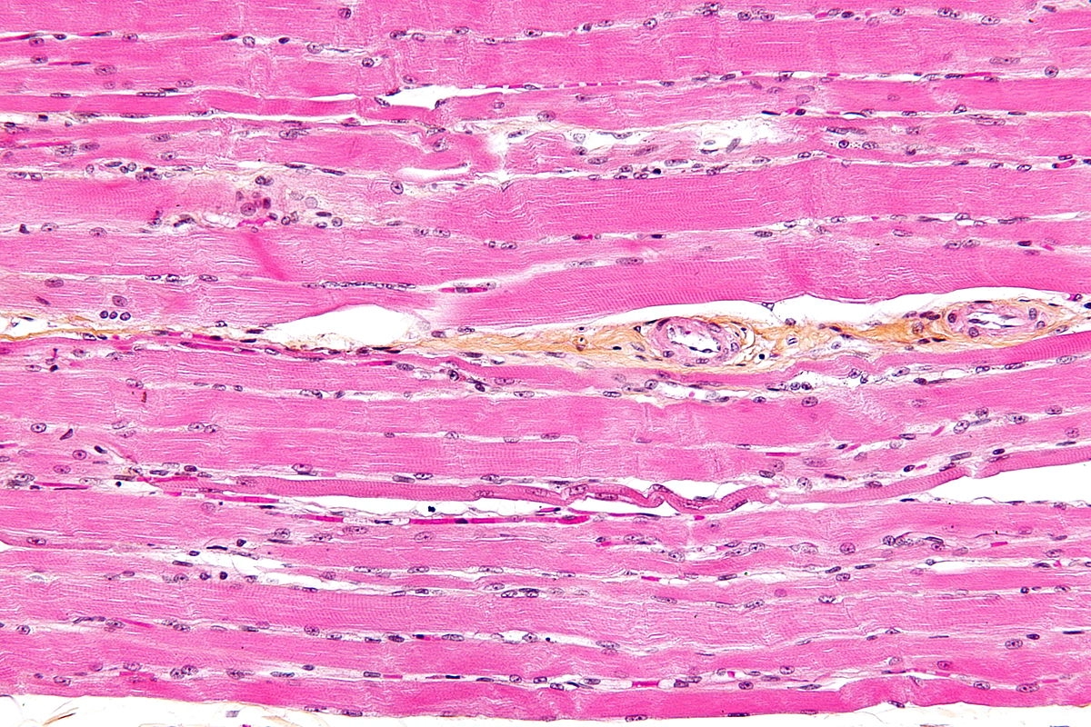
Skeletal striated muscle as seen under the microscope, after staining Image by Nephron, CC BY-SA 3.0, via Wikimedia Commons
-
Striated muscle is made up of muscle fibres
-
A muscle fibre is a highly specialised cell-like unit:
- Each muscle fibre contains an organised arrangement of contractile proteins in the cytoplasm
- Each muscle fibre is surrounded by a cell surface membrane
- Each muscle fibre contains many nuclei—this is why muscle fibres are not usually referred to as cells
-
The different parts of a muscle fibre have different names from the equivalent parts of a normal cell:
- Cell surface membrane = sarcolemma
- Cytoplasm = sarcoplasm
-
The sarcoplasm contains mitochondria and myofibrils
- The mitochondria carry out aerobic respiration to generate the ATP required for muscle contraction
- Myofibrils are bundles of actin and myosin filaments, which slide past each other during muscle contraction
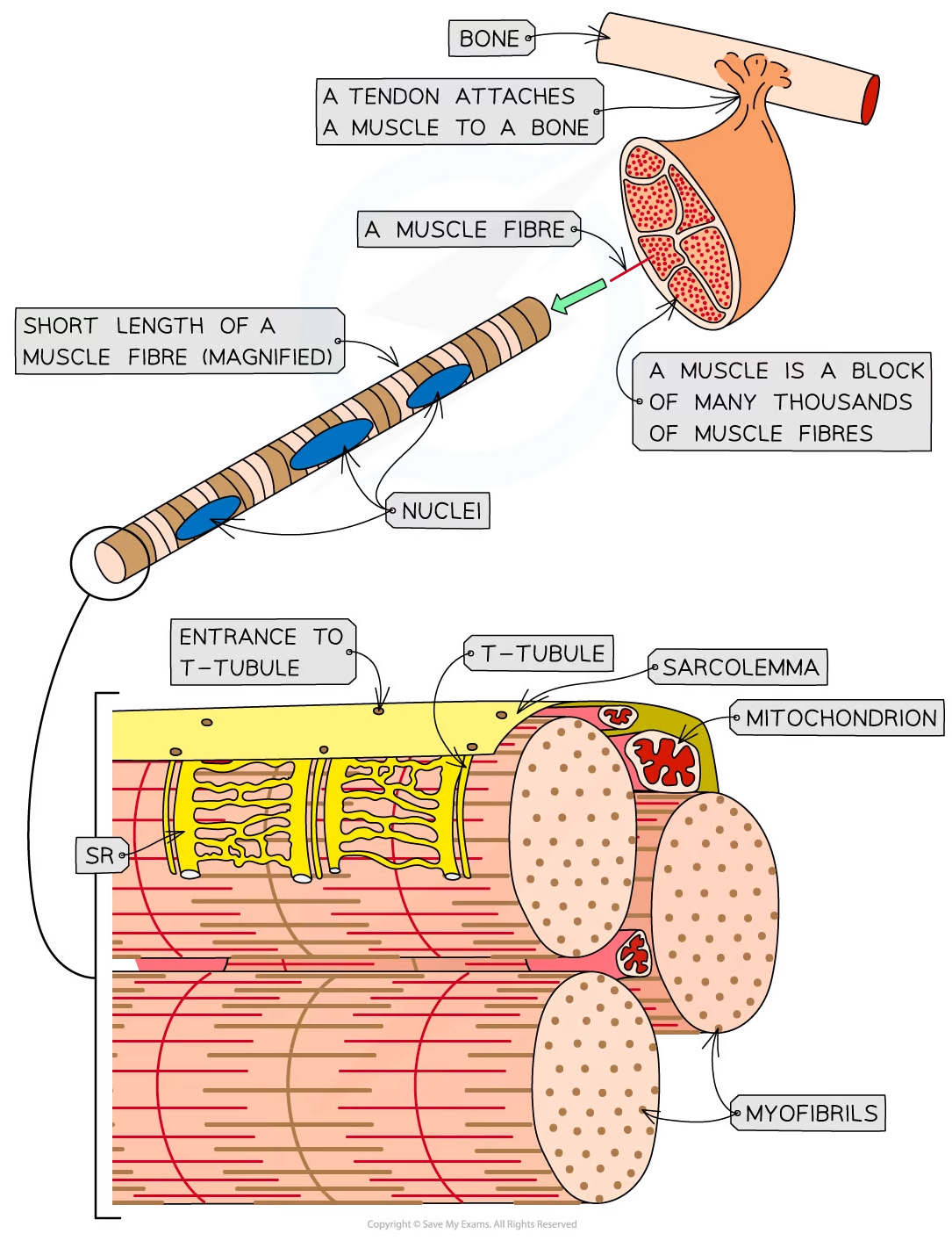
The ultrastructure of striated muscle and of a section of muscle fibre
Myofibrils
-
Myofibrils are located in the sarcoplasm
-
Each myofibril is made up of two types of protein filament:
- Thick filaments made of myosin
- Thin filaments made of actin
-
These two types of filament are arranged in a particular order, creating different types of band and line
| Part of myofibril | Description |
|---|---|
| H band | Only thick myosin filaments present |
| I band | Only thin actin filaments present |
| A band | Contains areas where only myosin filaments are present and areas where myosin and actin filaments overlap |
| M Line | Attachment for myosin filaments |
| Z line | Attachment for actin filaments |
| Sarcomere | The section of myofibril between two Z lines |
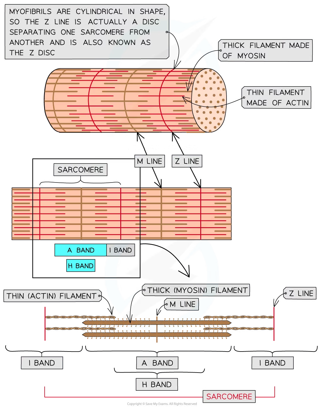
The structure of a myofibril
Was this revision note helpful?