Electron Microscopy of Plant Cells (Edexcel IAL Biology)
Exam board Pearson Edexcel · IALUpdated 2026-07-02
Spec point —
4.2
Electron Microscopy of Plant Cells
-
It is important to be able to recognise various plant organelles from electron microscope images
-
Chloroplast
- Has distinctive stacks of thylakoids
- Double membrane
- Has a roughly oval shape
- Larger than mitochondria
- Indicates the cell is a plant cell
-
Nucleus
- Has a nuclear membrane and a dark nucleolus within
- It has a roughly spherical shape
-
Vacuole
- Occupies a large space within a cell
- Often shows up as a very light shade (white) within an electron micrograph
- Indicates the cell is a plant cell
-
Cell wall
- Located around the perimeter of the cell
-
Mitochondria
- Roughly oval-shaped
- Double membrane
- Sometimes observed with visible cristae(foldings of the inner membrane)
-
Cell membrane (plasma membrane)
- Located just inside the cell wall, pressed against it
- Appears as a thin, dark line following the inner edge of the cell wall
- Often difficult to distinguish from the cell wall at low magnification, as the two run parallel and close together
- Encloses the cytoplasm and all the cell's content
-
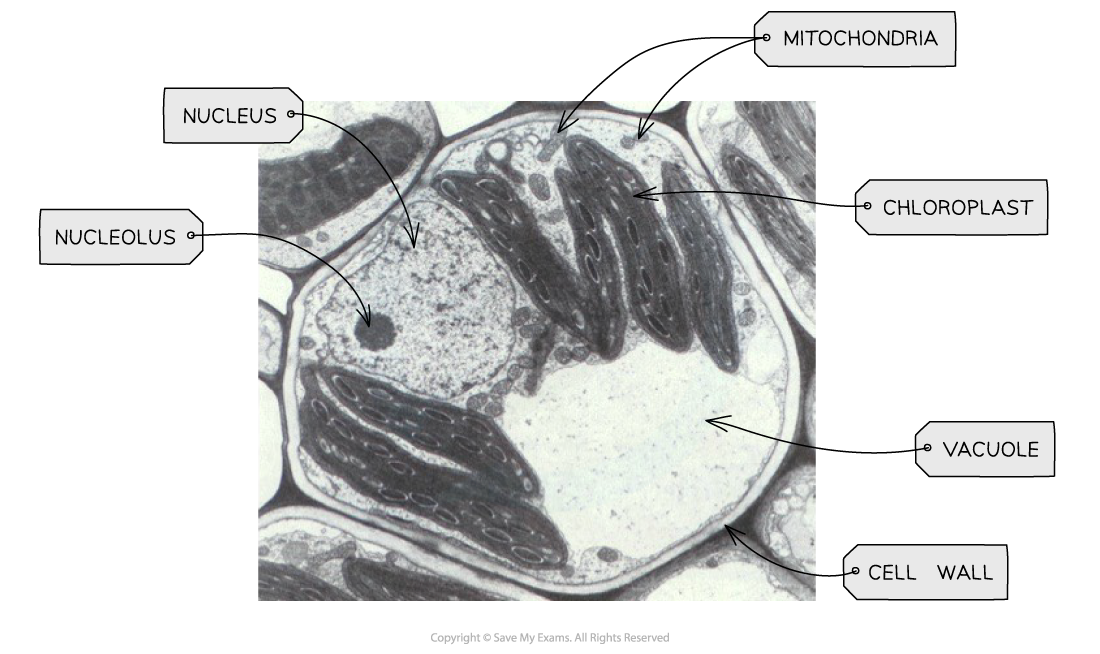
TEM electron micrograph of a plant cell showing key features
Was this revision note helpful?