Core Practical 7: Identifying Tissue Types Within Stems (Edexcel IAL Biology)
Exam board Pearson Edexcel · IALUpdated 2026-07-02
Identifying Tissue Types Within Stems
Spec point —
4.6
- In order to identify tissue types within stems, a permanent pre-prepared slide could be used
- Alternatively, a section of a plant stem could be cut and stained before preparing a temporary slide
Apparatus
- Plant stem
- Scalpel
- Suitable stain
- Microscope slide
- Cover slip
- Forceps
- Dissecting needle
- Light microscope
- Gloves
Method
- Cut a very thin cross-section of the stem using a scalpel
- Carefully transfer each section into a dish containing a suitable stain and leave for one minute
- E.g. toluidine blue O (TBO) stains xylem and sclerenchyma fibres blue-green, and phloem will appear pinkish purple
- Rinse off each section in water and mount onto a microscope slide, before adding a cover slip
- take care to lower the coverslip slowly over the sample from one side to the other to avoid trapping air bubbles, which could be mistaken for plant tissue structures
- View under a microscope and adjust the focus to form a clear image
- Make a labelled drawing of the positions of the xylem vessels, phloem sieve tubes and sclerenchyma fibres
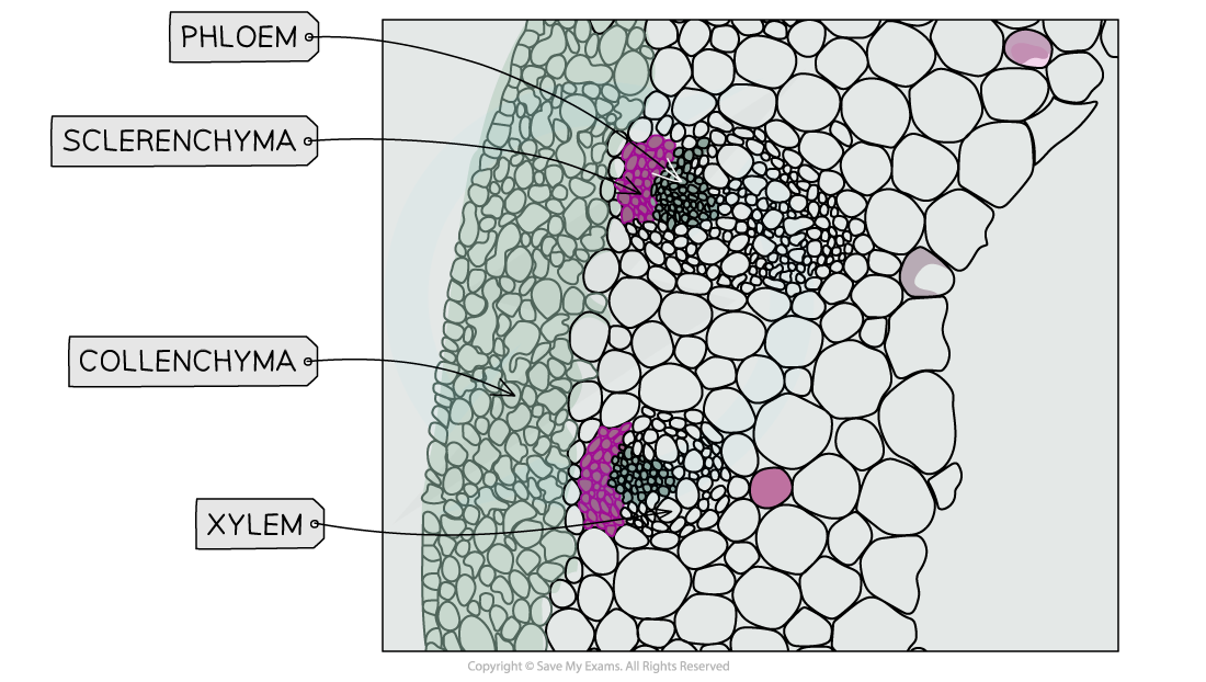
Microscopes can be used to view the location of different plant tissues within a cross-section; note that the stain used to generate this image is not the TBO stain described in the method above
Plan diagrams
-
When drawing tissue plan diagrams you need to:
- Read the instructions carefully
- Draw a large diagram
- Use a sharp pencil and do not shade (including the nucleus)
- Use clear, continuous lines (no sketching)
- When using an eyepiece graticule, use it to ensure you have correct proportions or if you are not using a microscope then endeavour to keep the proportions between tissues to scale
-
If drawing from a low-power image:
- Do not draw individual cells
- Read the question carefully as you may only have to draw a portion of the image
- Include the magnification on the drawing
-
If drawing from a high-power image:
- Draw only a few of the required cells
- Draw the cell wall of the plant cells
- Include the magnification on the drawing
-
When labelling, remember:
- Use a ruler for label lines (and scale lines if appropriate)
- Label lines should stop exactly at the structure (do not use arrows)
- Don't cross label lines over each other
- Label all tissues and relevant structures that are requested
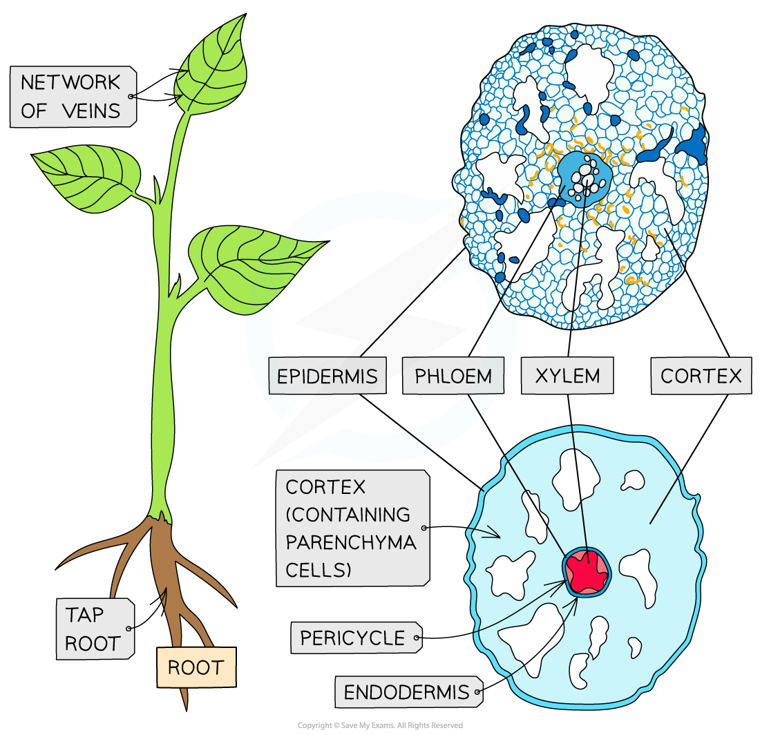
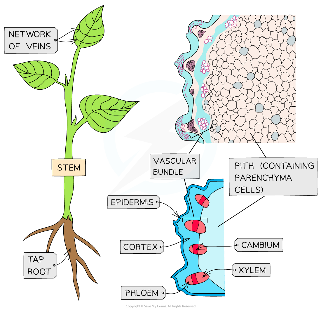
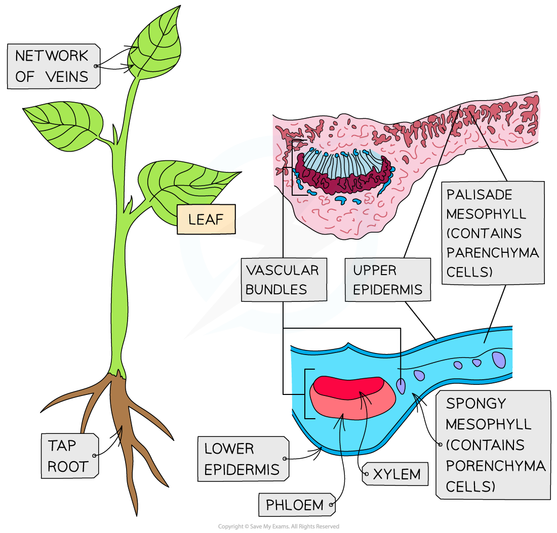
Tissue plan drawings can be produced from cross-sections of roots (top), stems (middle) and leaves (bottom)
Was this revision note helpful?