The Oxygen Dissociation Curve (Cambridge International AS Biology)
Spec point —
8.2.4· describe and explain the oxygen dissociation curve of adult haemoglobin
The oxygen dissociation curve
-
The oxygen dissociation curve shows the rate at which oxygen associates and dissociates with haemoglobin at different partial pressures of oxygen (pO2)
-
Partial pressure of oxygen refers to the pressure exerted by oxygen within a mixture of gases
- It is a measure of oxygen concentration
-
Haemoglobin is referred to as being saturated when all of its oxygen binding sites are taken up with oxygen
- This is when it contains four oxygen molecules
-
-
The ease with which haemoglobin binds and dissociates with oxygen can be described as its affinity for oxygen
- When haemoglobin has a high affinity it binds easily and dissociates slowly
- When haemoglobin has a low affinity for oxygen it **binds slowly **and dissociates easily
-
In other liquids, such as water, we would expect oxygen to become associated with water, or to dissolve, at a constant rate, providing a straight line on a graph
-
However, with haemoglobin, oxygen binds at different rates as the pO2** changes**
- This results in a curve when plotted
- It can be said that haemoglobin's affinity for oxygen changes at different partial pressures of oxygen
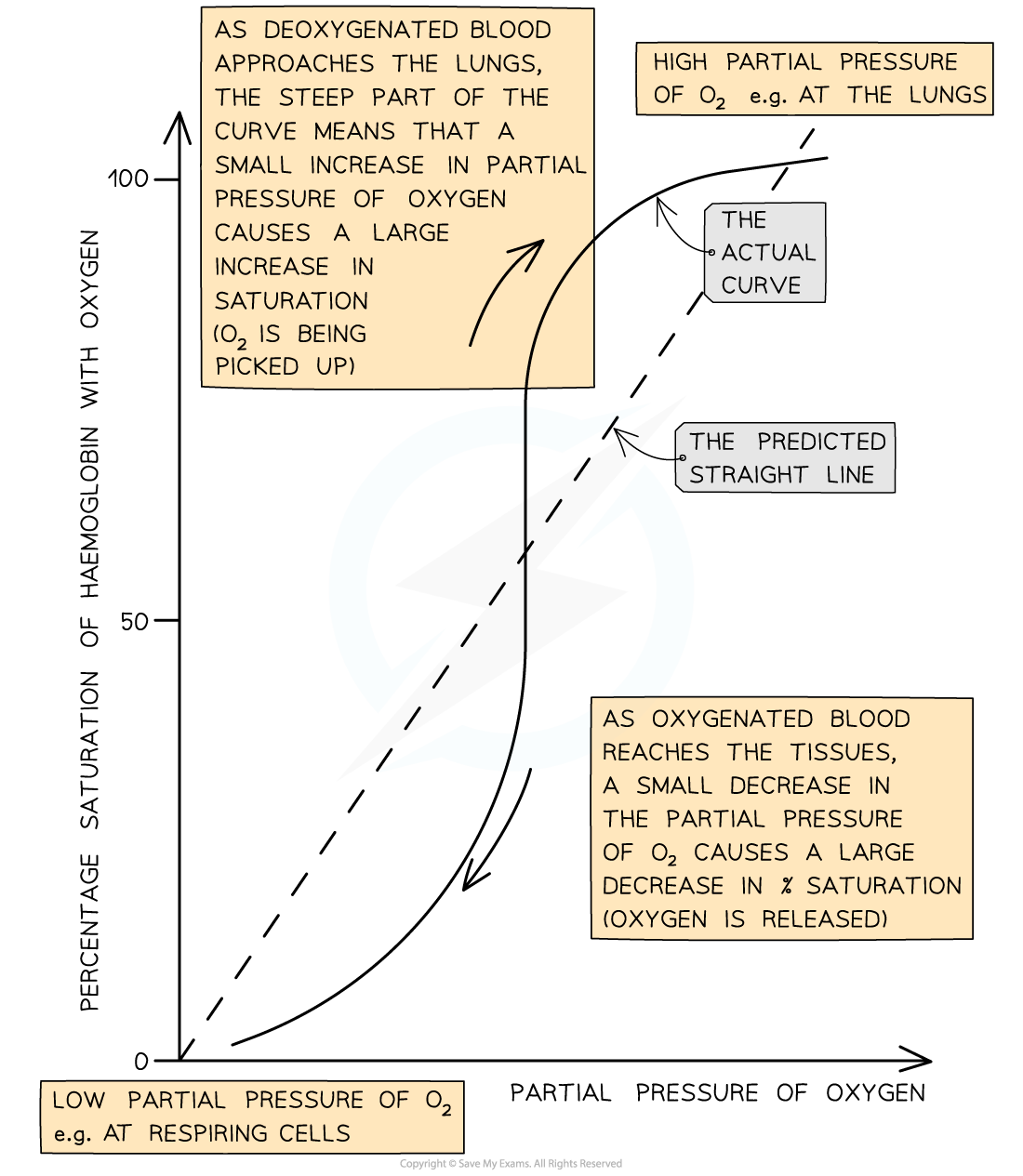
The oxygen dissociation curve is a sigmoidal shape
Interpreting the curve
-
When the curve is read from left to right, it provides information about the rate at which haemoglobin binds to oxygen at different partial pressures of oxygen
-
At low pO2 (in the bottom left corner of the graph) oxygen binds slowly to haemoglobin
-
This means that haemoglobin cannot pick up oxygen and become saturated as blood passes through the body's oxygen-depleted tissues
- Haemoglobin has a low affinity for oxygen at low pO2, so saturation percentage is low
-
At medium pO2 (in the central region of the graph) oxygen binds more easily to haemoglobin and saturation increases quickly
- At this point on the graph a small increase in pO2causes a large increase in haemoglobin saturation
-
At high pO2 (in the top right corner of the graph) oxygen binds easily to haemoglobin
-
This means that haemoglobin can pick up oxygen and become saturated as blood passes through the lungs
- Haemoglobin has a high affinity for oxygen at high pO2**, **so saturation percentage is high
- Note that at this point on the graph, increasing the pO2** by a large amount only has a small effect on the percentage saturation of haemoglobin**
- This is because most oxygen binding sites on haemoglobin are already occupied
-
-
When read from right to left, the curve provides information about the rate at which haemoglobin dissociates with oxygen at different partial pressures of oxygen
-
In the lungs, where pO2is high
- There is very little dissociation of oxygen from haemoglobin
-
At medium pO2, oxygen dissociates readily from haemoglobin, as shown by the steep region of the curve
- This region corresponds with the partial pressures of oxygen present in the respiring tissues of the body, so release of oxygen is important for cellular respiration
- At this point on the graph a small decrease in pO2causes a large decrease in percentage saturation of haemoglobin, leading to easy release of plenty of oxygen to the cells
-
At low pO2 dissociation slows again
- There are few oxygen molecules left on the binding sites, and the release of the final oxygen molecule becomes more difficult, in a similar way to the slow binding of the first oxygen molecule
-
Explaining the Oxygen Dissociation Curve
Spec point —
8.2.5· explain the importance of the oxygen dissociation curve at partial pressures of oxygen in the lungs and in respiring tissues
-
The curved shape of the oxygen dissociation curve for haemoglobin can be explained as follows
-
Due to the shape of the haemoglobin molecule it is difficult for the first oxygen molecule to bind to haemoglobin
- This means that binding of the first oxygen occurs slowly
- This explains the relatively shallow curve at the bottom left corner of the graph
-
After the first oxygen molecule binds to haemoglobin, the haemoglobin protein changes shape, or conformation
- This makes it easier for the next oxygen molecules to bind
- This speeds up binding of the remaining oxygen molecules and explains the steeper part of the curve in the middle of the graph
- The shape change of haemoglobin leading to easier oxygen binding is known as cooperative binding
-
As the haemoglobin molecule approaches saturation it takes longer for the fourth oxygen molecule to bind due to the shortage of remaining binding sites
- This explains the levelling off of the curve in the top right corner of the graph
-
Was this revision note helpful?
Build on this topic